Fatty liver (MASLD) and abnormal liver tests
Understanding raised liver enzymes, the FIB-4 score, elastography and liver fibrosis
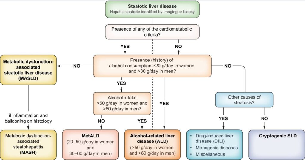
Fatty liver disease is now the most common cause of abnormal liver tests in Australia. The condition previously known as Non-Alcoholic Fatty Liver Disease (NAFLD), has been renamed Metabolic Dysfunction-Associated Steatotic Liver Disease (MASLD). This change reflects our improved understanding that liver fat is usually driven by metabolic factors such as obesity, type 2 diabetes, insulin resistance and central adiposity.
Why liver scarring matters more than liver fat
The good news is that most people with MASLD will never develop serious liver disease. However, the challenge is identifying the minority who develop significant liver scarring, also called fibrosis. This matters because fibrosis, rather than liver fat itself, is what best predicts future risks of cirrhosis, liver failure, liver cancer and liver-related death.
10 key pearls about fatty liver and abnormal liver tests
GP quick reference
FIB-4
25–29.9 — overweight
30–34.9 — obesity class I
35–39.9 — obesity class II
≥40 — obesity class III / severe
BMI ≥25 kg/m²; use ≥23 kg/m² for Asian ancestry
Waist ≥80 cm women; ≥94 cm men; ≥90 cm Asian men
Fasting glucose ≥5.6 mmol/L, HbA1c ≥5.7%, T2DM or treatment
BP ≥130/85 mmHg or antihypertensive treatment
Triglycerides ≥1.7 mmol/L or treatment
HDL ≤1.0 mmol/L men / ≤1.3 mmol/L women or treatment
Women: MASLD range <140 · MetALD 140–350 · ALD range >350 g/week
Men: MASLD range <210 · MetALD 210–420 · ALD range >420 g/week
<0.05 µmol/L — no/light recent alcohol exposure
0.05–<0.28 µmol/L — emerging PEth-MetALD range
≥0.28 µmol/L — emerging PEth-ALD range
PEth reflects alcohol exposure over approximately 2–4 weeks. Interpret with quantified alcohol history (g/week) ± AUDIT-C. Current thresholds are emerging, not formal diagnostic criteria.
✓ Interpret risk and next steps
✓ Includes PEth (µmol/L) guidance
✓ Alcohol-aware elastography interpretation
Important: FIB-4 should always be interpreted in clinical context. A low score reduces the likelihood of advanced fibrosis, but it does not completely exclude it, particularly in younger patients or those with significant metabolic risk factors.
FIB-4 + elastography calculator
For clinical use. FIB-4 = (Age × AST) ÷ (Platelets × √ALT)
Women: MASLD range <140 · MetALD 140–350 · ALD range >350 g/week
Men: MASLD range <210 · MetALD 210–420 · ALD range >420 g/week
<0.05 µmol/L — no/light recent alcohol exposure
0.05–<0.28 µmol/L — emerging PEth-MetALD range
≥0.28 µmol/L — emerging PEth-ALD range
A practical first step
Abnormal liver tests should usually be confirmed with repeat testing and assessment for common causes. Useful investigations often include:
Normal liver tests do not exclude silent liver disease
Many patients with significant liver fibrosis — and even some with cirrhosis — have liver enzyme levels within the laboratory reference range.
The absence of an elevated ALT does not exclude clinically important silent liver disease.
What is a normal ALT?
Traditional laboratory reference ranges often define normal ALT as <40 U/L in men and <30 U/L in women. However, healthy population studies suggest the true upper limits of normal are lower:
ALT values above these levels warrant consideration of underlying liver disease, particularly in patients with obesity, type 2 diabetes or metabolic syndrome.
Alcohol history
Accurate alcohol assessment is important because metabolic fatty liver disease, MetALD and alcohol-related liver disease can overlap. Intake is often underestimated — a written or electronic questionnaire may be more accurate than verbal questioning.
Rather than simply asking "How much do you drink?", more useful questions include:
One or more such episodes suggests higher-risk alcohol use and should prompt more detailed assessment. Alcohol questions should be asked routinely and non-judgementally as part of standard liver health assessment.
If alcohol is contributing
Reducing or stopping alcohol can significantly improve liver health. However, people who drink heavily every day should not stop abruptly without medical advice — alcohol withdrawal can cause seizures, delirium and, rarely, death.
Management may include:
Alcohol use disorder is a medical condition, and effective treatments are available.
Supplements and herbal products
Many patients do not consider supplements, herbal remedies or "natural" products to be medications and may not mention them unless specifically asked.
An important point
The highest-risk products are often multi-ingredient supplements marketed for weight loss, bodybuilding, energy, detoxification or general wellness. These may contain numerous active ingredients, variable concentrations, contaminants or ingredients not clearly listed on the label.
Garcinia cambogia
Detox teas
Liver cleanse products
Weight loss supplements online
Testosterone boosters
SARMs
Fat burners
Pre-workout supplements
Kava
Kratom
Herbal sleep aids
Calming supplements
Menopause supplements
Fertility supplements
Hormonal support products
High-dose niacin
Cholesterol-lowering supplements
Herbal anti-inflammatories
Multi-ingredient wellness supplements
Ayurvedic medicines
He Shou Wu (Polygonum multiflorum)
Herbal mixtures from overseas
Recognising supplement-induced liver injury
Supplement-related liver injury is often overlooked because patients may not consider herbal or "natural" products to be medications. Clues include:
The culprit is often a multi-ingredient product, making identification difficult. When unexplained liver test abnormalities are identified, stopping all non-essential supplements is often an important first step while further assessment is undertaken.
Drug-induced liver injury (DILI)
Many medications can affect liver tests, although true drug-induced liver injury is relatively uncommon. When abnormal liver tests are identified, it is important to review all prescription medications, over-the-counter products, supplements and complementary medicines.
Medications most commonly considered include:
Although commonly reviewed, clinically significant liver injury from these medications remains uncommon.
A note on statins
Statins frequently cause mild elevations in liver enzymes but rarely cause clinically significant liver injury. Statins are safe and beneficial in most patients with MASLD and should not be routinely discontinued simply because fatty liver or mildly abnormal liver tests are present.
Example
A patient with established ischaemic heart disease starts rosuvastatin 20 mg and ezetimibe 10 mg. Their ALT rises from 100 to 220 U/L, then stabilises at 150–200 U/L. Bilirubin, INR, albumin and platelets remain normal. Fibrosis assessment remains reassuring. In this situation, continuing lipid-lowering therapy with monitoring may be safer than stopping effective cardiovascular protection because of the ALT alone.
The decision should be based on the whole clinical picture — not the ALT number alone.
Medications that can mimic or worsen fatty liver disease
These warrant particular attention in patients with obesity, diabetes, metabolic syndrome or pre-existing liver disease:
Clues suggesting DILI
Drug-induced liver injury should be considered when:
The timing can be deceptive — drug-induced liver injury often occurs several weeks after a medication or supplement is started.
Practical approach
The decision to stop a medication should always balance potential liver risk against the benefit of treatment. Questions worth asking include:
Not every abnormal liver test is drug-induced liver injury, and not every potentially hepatotoxic medication needs to be stopped.
Understanding liver fibrosis
Fibrosis refers to scarring within the liver. The stage of fibrosis — not the amount of fat — is what determines long-term risk.
Advanced fibrosis (F3) and cirrhosis (F4) are associated with increased risks of liver failure, portal hypertension, liver cancer and liver-related death.
The encouraging news: fibrosis, particularly F3, may improve with weight loss, better metabolic health and treatment of contributing factors.
How common is MASLD?
Among adults over 50 years of age:
Risk factors include:
Waist circumference above 80 cm in women or 94 cm in men suggests increased metabolic risk.
Diagnosing MASLD
Diagnosis involves excluding other causes of liver disease and assessing fibrosis risk. Blood tests are used to exclude hepatitis B, hepatitis C, haemochromatosis, autoimmune liver disease and drug-induced liver injury.
Ultrasound is useful for detecting liver fat but cannot reliably determine the degree of fibrosis. A normal ultrasound does not exclude liver fibrosis.
The FIB-4 score
The FIB-4 score estimates the risk of advanced liver fibrosis using age, AST, ALT and platelet count. It is the recommended first-line fibrosis assessment tool.
Patients aged 65 and over: some guidelines recommend a higher low-risk threshold of approximately 1.9–2.0, as age can inflate the score.
Liver elastography (FibroScan®)
Elastography measures liver stiffness, which correlates with the degree of fibrosis.
Treatment of MASLD
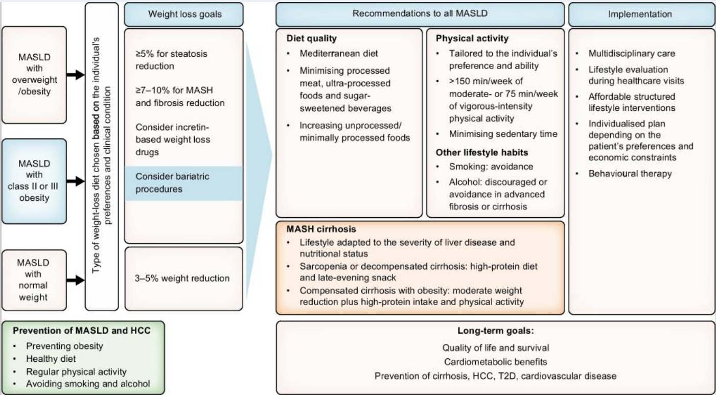
MASLD sits at the intersection of liver disease and metabolic syndrome. The most effective treatment targets the underlying drivers rather than the liver in isolation. For most patients, there is no single "liver medication" that replaces weight loss, metabolic health and cardiovascular risk reduction.
Coffee
Regular coffee consumption has been associated with lower rates of fatty liver, liver fibrosis, cirrhosis and liver cancer — across MASLD, alcohol-related liver disease and viral hepatitis. It is one of the few simple dietary habits consistently associated with better liver outcomes.
Coffee is not a substitute for diet, weight loss, exercise, alcohol reduction or metabolic risk management — but for patients who already drink it, there is no liver-related reason to stop.
Newer therapies
Advanced fibrosis (F3–F4)
Patients with advanced fibrosis or cirrhosis require specialist assessment and structured surveillance. This may include:
Management of weight, diabetes, cholesterol, alcohol and cardiovascular risk remains important even after advanced fibrosis has developed.
Can liver fibrosis improve?
Yes. Unlike many forms of organ scarring, liver fibrosis can improve when the underlying cause is treated.
This is one of the reasons early detection matters — intervention can meaningfully alter long-term outcomes.
Key messages
Understanding Liver Fibrosis
Fibrosis refers to scarring within the liver.
| Stage | Meaning |
|---|---|
| F0 | No fibrosis |
| F1 | Mild fibrosis |
| F2 | Moderate fibrosis |
| F3 | Advanced fibrosis |
| F4 | Cirrhosis |
Why Fibrosis Matters
The most important question is not whether fat is present in the liver, but whether significant fibrosis has developed.
Advanced fibrosis (F3) and cirrhosis (F4) are associated with increased risks of:
- Liver failure
- Portal hypertension
- Liver cancer (hepatocellular carcinoma)
- Liver-related mortality
The encouraging news is that fibrosis, particularly F3 fibrosis, may improve with weight loss, improved metabolic health and treatment of contributing factors.
Fatty Liver Disease (MASLD)
The New Terminology
MASLD stands for Metabolic Dysfunction-Associated Steatotic Liver Disease.
How Common Is MASLD?
Among adults over 50 years of age:
- Approximately 1 in 2 have steatotic liver disease
- Up to 9 in 10 people with obesity, type 2 diabetes or metabolic syndrome have liver fat
- Many people have completely normal liver tests despite significant liver disease
Who Is at Risk?
Risk factors include:
- Obesity
- Type 2 diabetes
- Prediabetes
- Hypertension
- Elevated triglycerides
- Metabolic syndrome
- Central abdominal obesity
A waist circumference above approximately:
- 80 cm in women
- 94 cm in men
suggests increased metabolic risk.
Diagnosing MASLD
Diagnosis usually involves excluding other causes of liver disease and assessing fibrosis risk.
Blood Tests
To exclude:
- Hepatitis B
- Hepatitis C
- Haemochromatosis
- Autoimmune liver disease
- Drug-induced liver injury
Ultrasound
Ultrasound is useful for detecting liver fat but cannot reliably determine the degree of fibrosis.
A normal ultrasound does not exclude liver fibrosis.
The FIB-4 Score
The FIB-4 score estimates the risk of advanced liver fibrosis using:
- Age
- AST
- ALT
- Platelet count
Interpreting the FIB-4 Score
| FIB-4 Score | Risk of Advanced Fibrosis | Suggested Next Step |
|---|---|---|
| <1.3 | Low risk | Lifestyle and metabolic risk factor management |
| 1.3-2.67 | Indeterminate risk | Liver elastography |
| >2.67 | High risk | Specialist assessment |
Patients Aged 65 Years and Older
Some guidelines recommend a higher low-risk threshold of approximately 1.9-2.0 in patients aged 65 years and over.
Liver Elastography (FibroScan®)
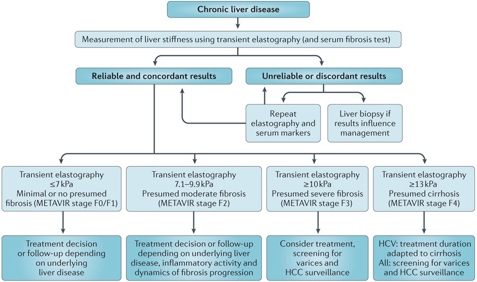
Elastography measures liver stiffness, which correlates with fibrosis.
| Liver Stiffness Measurement | Interpretation |
|---|---|
| <8 kPa | Advanced fibrosis unlikely |
| 8-12 kPa | Possible significant fibrosis |
| >12 kPa | Advanced fibrosis or cirrhosis increasingly likely |
Treatment of MASLD
MASLD sits at the intersection of liver disease and metabolic syndrome. The liver is injured by metabolic dysfunction, so the most effective treatment targets the underlying drivers rather than the liver in isolation.
For most patients, there is no single "liver medication" that replaces weight loss, metabolic health and cardiovascular risk reduction.
The Core Treatments
Weight Loss
- A weight loss of 5-7% reduces liver fat
- A weight loss of 10% or more may improve steatohepatitis (MASH)
- Early fibrosis may stabilise or regress with sustained weight loss
Physical Activity
Exercise reduces liver fat independently of weight loss.
Aim for at least 150 minutes of moderate physical activity per week.
Diabetes and Insulin Resistance
Improving glucose control can improve liver health.
GLP-1 receptor agonists such as semaglutide and tirzepatide appear to provide liver benefits beyond their effects on weight and blood sugar control.
Cholesterol and Triglycerides
Statins are safe in MASLD and are frequently underused because of concerns about liver toxicity.
Cardiovascular disease remains the leading cause of death in patients with MASLD.
Blood Pressure and Sleep Apnoea
Hypertension and obstructive sleep apnoea commonly coexist with MASLD and should be actively identified and treated.
Alcohol
Alcohol and metabolic dysfunction can act together to accelerate liver injury.
Even modest alcohol intake may accelerate fibrosis progression in some patients with MASLD, particularly when significant fibrosis is already present.
Medications and Supplements
Some medications can worsen steatosis or steatohepatitis, including corticosteroids, tamoxifen, amiodarone and methotrexate.
Supplements marketed for liver health, detoxification, weight loss or bodybuilding may themselves cause liver injury and should always be reviewed.
Coffee
Regular coffee consumption has been associated with lower rates of fatty liver, liver fibrosis, cirrhosis and liver cancer.
The benefit appears to extend across several liver diseases, including MASLD, alcohol-related liver disease and viral hepatitis.
For patients who already drink coffee, there is no liver-related reason to stop.
For patients who tolerate coffee, around 2-4 cups per day may be reasonable as part of a liver-health plan.
The brewing method probably matters less than is sometimes suggested. Most of the evidence showing liver benefit does not clearly distinguish between espresso, filtered coffee, instant coffee or plunger coffee.
A practical approach is:
- Drink coffee without added sugar, syrups or cream
- Choose the style of coffee you tolerate and enjoy
- Consider filtered coffee if LDL cholesterol is difficult to control
- Limit caffeine if it worsens insomnia, anxiety, reflux, palpitations or blood pressure
Unfiltered coffee, including plunger/French press coffee, may raise LDL cholesterol in some people. Espresso is technically unfiltered, but the serving size is smaller and the clinical impact is uncertain.
In patients who need strong cardiovascular prevention, the answer is not usually to stop coffee; it is to manage cardiovascular risk properly, including LDL cholesterol when indicated.
Coffee is not a substitute for diet, weight loss, exercise, alcohol reduction or metabolic risk management, but it is one of the few simple dietary habits consistently associated with better liver outcomes.
Newer Therapies
GLP-1 receptor agonists such as semaglutide and tirzepatide have demonstrated improvements in liver inflammation and fibrosis in clinical trials.
Vitamin E (800 IU daily) has been shown to improve steatohepatitis in selected patients with biopsy-proven MASH who do not have diabetes. It has not consistently been shown to improve fibrosis and is generally considered on an individual basis rather than routinely recommended for all patients.
Resmetirom became the first medication specifically approved for MASH with fibrosis in 2024. Availability varies between countries but it represents an important advance in targeted therapy for patients with significant fibrosis.
Advanced Fibrosis (F3-F4)
Patients with advanced fibrosis or cirrhosis require specialist assessment and structured surveillance.
This may include:
- Six-monthly liver ultrasound
- Alpha-fetoprotein (AFP) monitoring
- Assessment for oesophageal varices
- Monitoring for portal hypertension and liver failure
Importantly, management of weight, diabetes, cholesterol, alcohol intake and cardiovascular risk remains beneficial even after advanced fibrosis has developed.
The Most Important Question
When fatty liver disease is identified, the key question is not:
"Is there fat in the liver?"
The key question is:
"Is there advanced fibrosis?"
Fat in the liver is extremely common.
Advanced fibrosis is much less common, but it is fibrosis that predicts future risks of:
- Cirrhosis
- Liver failure
- Portal hypertension
- Liver cancer
- Liver-related mortality
Modern liver assessment therefore focuses on identifying the small proportion of patients with advanced fibrosis while avoiding unnecessary investigations in the much larger group with uncomplicated steatotic liver disease.
Can Liver Fibrosis Improve?
Yes.
Unlike many forms of organ scarring, liver fibrosis can improve when the underlying cause is treated.
Studies show:
- A weight loss of 5-7% improves liver fat accumulation
- A weight loss of 10% or more can resolve steatohepatitis (MASH) in many patients
- Advanced fibrosis (F3) may improve or even regress with sustained weight loss and metabolic control
This is one of the reasons early detection of fibrosis is so important: intervention can meaningfully alter long-term outcomes.
Key Messages
- MASLD is now the most common cause of abnormal liver tests in Australia.
- Normal liver tests do not exclude significant liver disease.
- Ultrasound detects liver fat but does not reliably assess fibrosis.
- The FIB-4 score is the recommended first-line fibrosis assessment tool.
- Elastography helps determine whether advanced fibrosis is present.
- The most important question is not whether fat is present, but whether fibrosis is present.
- Advanced fibrosis (F3-F4) warrants specialist assessment and ongoing monitoring.
- Weight loss, improved metabolic health and reduced alcohol intake can significantly improve liver outcomes.
- Liver fibrosis can improve, and sometimes regress, when the underlying cause is treated.
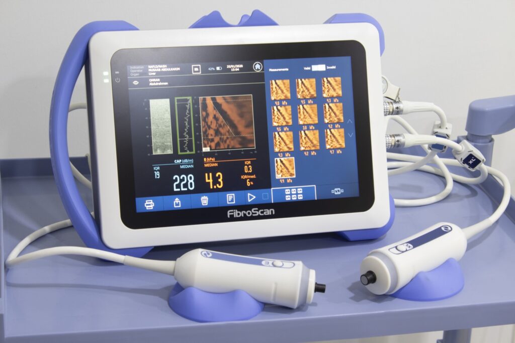
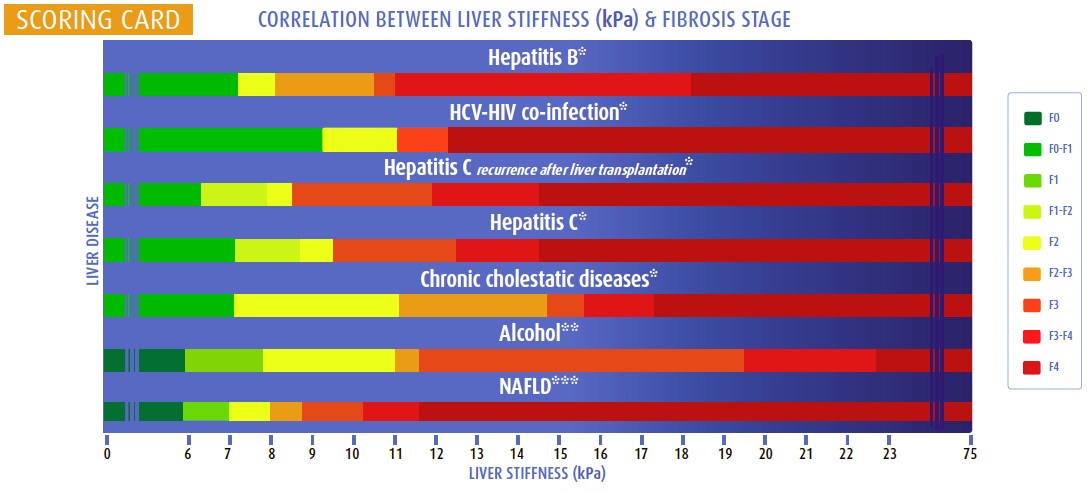
Liver Stiffness Measurements - FIBROSCAN
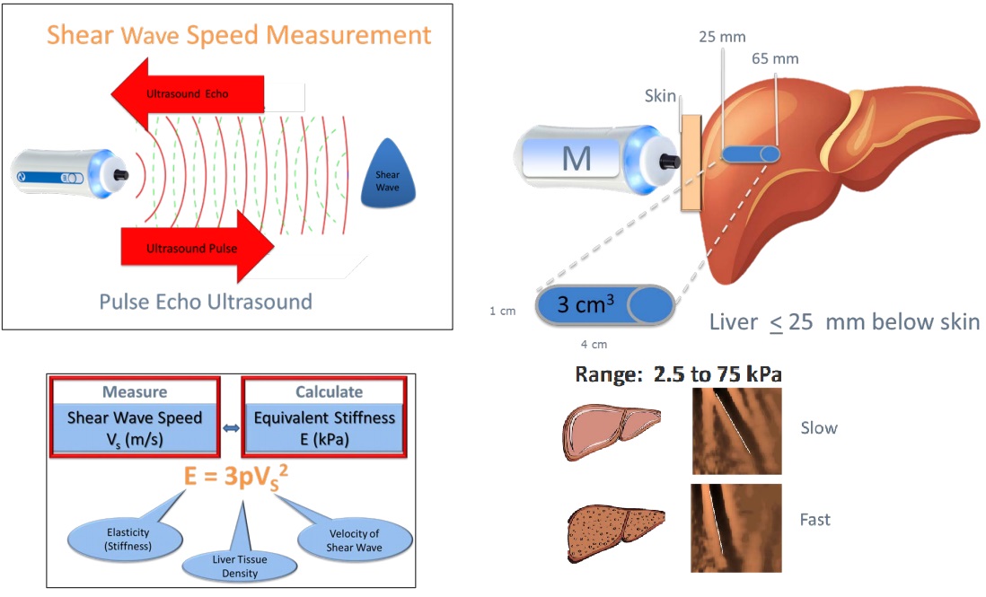
Important Differences Between SWE and Fibroscan (TE)
| Feature | Shear Wave Elastography (SWE) | Fibroscan (Transient Elastography, TE) |
|---|---|---|
| How shear wave is generated | Acoustic radiation force (ultrasound pulse) | Mechanical external pulse |
| Equipment | Standard ultrasound machine | Dedicated Fibroscan® device |
| Availability | Widely available | Specialist settings |
| Calibration | Machine-specific (some variability) | Highly standardised globally |
| Interpretation thresholds | Slightly higher readings than TE at low fibrosis stages | Standard fibrosis thresholds validated for TE |
General Interpretation of SWE and Fibroscan (TE) Readings
| SWE (kPa) | TE (kPa) | Interpretation (likely) | Suggested Action |
|---|---|---|---|
| <7.5 | <7.0 | No significant fibrosis | Routine monitoring |
| 7.5–8.0 | 7.0–8.0 | Borderline; mild fibrosis | Consider clinical context |
| >8.0–9.5 | >8.0–9.5 | Suspicious for fibrosis | Recommend confirmation with Fibroscan (TE) |
| >9.5–12.0 | >9.5–12.5 | Significant fibrosis | Fibroscan (TE) or specialist review recommended |
| >12.0 | >12.5 | Advanced fibrosis or cirrhosis | Specialist referral strongly recommended |
Further review may be appropriate when:
-
Rising SWE readings over time, even if liver enzymes remain stable.
-
SWE >7.5 kPa in a patient with:
-
Metabolic risk factors (eg, BMI >30, diabetes, dyslipidaemia).
-
History of alcohol use (>14 standard drinks/week for men; >7 for women).
-
New, persistent, or unexplained elevated liver enzymes:
-
ALT or AST >2 × ULN (Upper Limit of Normal; ULN) where AST 30 U/L (female), 20 U/L (male)).
-
GGT >2 × ULN.
-
-
Practical guidance:
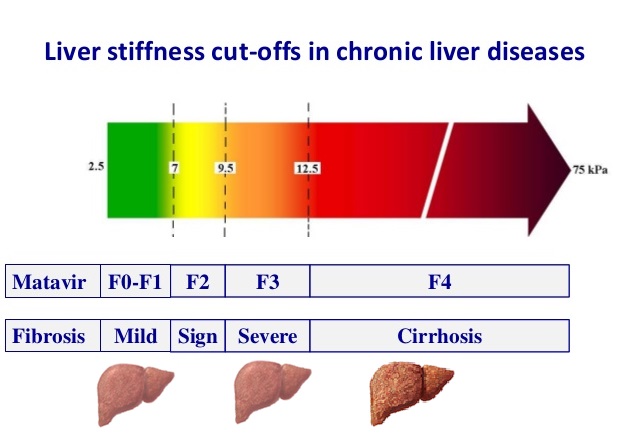
The below colour charts refer to Fibroscan (VCTE) not ultrasound Shearwave (SWE). If SWE >8.0 kPa and management decisions depend on accurate fibrosis staging, confirm with Fibroscan (VCTE) where available. When reading SWE reports: Confirm IQR/Median <30%, success rate >60%, and good technical quality comment.
clinically significant portal hypertension (CSPH): VCTE ≤15 kPa + platelets ≥150×109/l rules out CSPH in adults with MASLD; With compensated advanced chronic liver disease, LSM ≥20 kPa or platelets <150×109/l = varices screening/upper GI endoscopy; LSM ≥25 kPa rules in CSPH in non-obese (BMI <30 kg/m2) adults with MASLD; while obesity confounds LSM, no optimal non-invasive test to rule in CSPH in MASLD+obesity
Liver Scores and Biomarkers
Blood-based scores improve fibrosis assessment, especially when combined with elastography:
- APRI (AST to Platelet Ratio Index)
- FIB-4 (Age, AST, ALT, platelets)
- ELF (Enhanced Liver Fibrosis Score: TIMP-1, PIIINP, HA)
- FibroTest (α2-macroglobulin, haptoglobin, apolipoprotein A1, bilirubin, GGT)
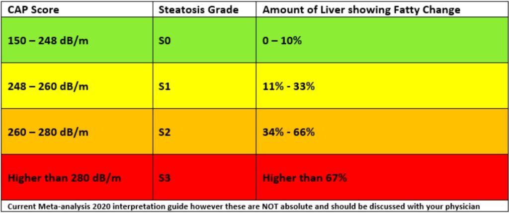
Liver Fat (CAP) Score
Fat accumulation (steatosis) is assessed non-invasively using Fibroscan's Controlled Attenuation Parameter (CAP):
- Measured in decibels per meter (dB/m)
- Scores range from 100 to 400 dB/m
- Higher CAP scores reflect greater degrees of steatosis
Routine vs Expanded Liver Screening During Therapy
| Scenario | Tests Recommended |
|---|---|
| Routine Monitoring (stable patient, normal LFTs) |
|
| Expanded Testing (persistent abnormal LFTs, or clinical suspicion) |
|
Summary
- If LFTs are normal and Fibroscan <7.0 kPa → continue routine monitoring (LFTs 3–6 monthly; Fibroscan every 5 years).
- If LFTs are >2× ULN or Fibroscan ≥8.0–9.5 kPa → consider expanded testing and/or hepatology referral.
- Obesity (BMI >30), diabetes, heavy alcohol use (>14/week for men, >7/week for women), or new hepatomegaly on examination/imaging are clinical triggers to lower the threshold for Fibroscan or broader testing.
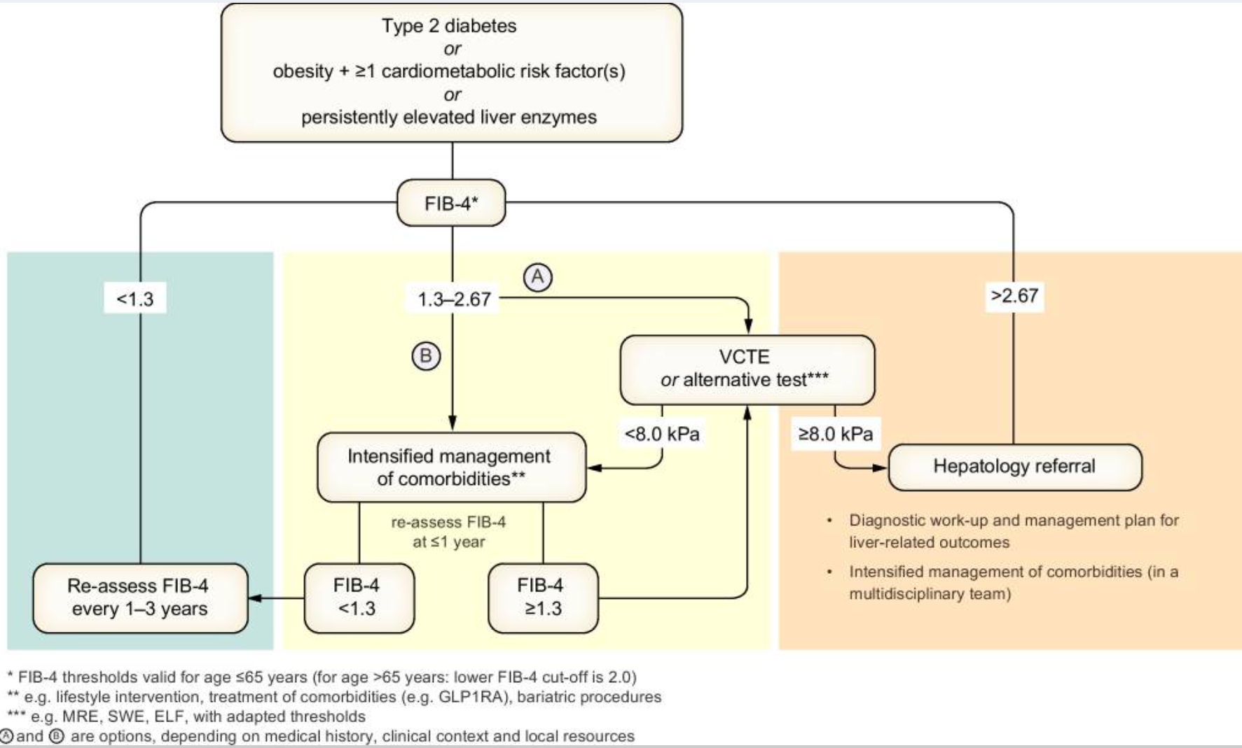
The FIB-4 Score: Fibrosis Risk in NAFLD
Formula:
(Age × AST) / (Platelets × √ALT) [Use online calculator – e.g. MDCalc]
FIB-4 Interpretation (age <65 years):
| FIB-4 Score | Fibrosis Risk | Action |
| <1.3* | Low | GP follow-up, order USS; optimise lifestyle |
| 1.3–2.67 | Intermediate | Order USS/FibroScan ± discuss with hepatology |
| >2.67 | High | Refer to hepatology |
*For age ≥65, <2.0 as the low-risk threshold
false positives: non-hepatic AST>ALT (exercise, meds, alcohol or muscle injury), low platelets (ITP, age), non-liver/fibrosis inflammation (infection/sepsis, autoimmunity, trauma/surgery)
false negatives: age<40, high metabolic burden/silent MASH-F3/4, CRP>5+ferritin>300 (F)/500(M) (ALT <30 F, <40 M - no inflammatory flare), Child's A cirrhosis +/- normal platelets, South+East Asia/Older adults
Summary:
positive predictive value (PPV) of FIB-4 >2.67 for advanced fibrosis (F3/4) varies by population and setting — particularly by prevalence of fibrosis.
| FIB-4 Cutoff | Setting | PPV for F3–4 |
|---|---|---|
| >2.67 | Primary care | ~60–70% |
| >2.67 | hepatology care | ~70–80% |
| >3.25 + Imaging | Combined strategy | ~90%+ |
In Primary Care Settings: PPV of FIB-4 >2.67 for F3/4 fibrosis is typically in the range of 50–70%, depending on population risk.
Lower end (~50%) in general adult screening populations.
Higher end (~70%) in enriched groups (e.g. T2DM, obesity, elevated ALT).
Management Plan in General Practice
First Steps:
- Address metabolic syndrome: weight loss (≥5–10%), diet, exercise
- 5–10% weight reduction = steatohepatitis+fibrosis improves
- >10% weight loss = steatohepatitis resolves in up to 90%
- Optimise diabetes, lipids, BP
- Avoid hepatotoxins (alcohol, excess paracetamol, unnecessary meds/supplements)
- repeat FIB-4 every 12-18 months
- Lifestyle + CVD risk reduction is the cornerstone
Refer if:
- FIB-4 >2.67 or abnormal FibroScan/SWE
- ALT persistently >2x ULN for >6 months (unexplained or higher risk false negative)
- Evidence of fibrosis, cirrhosis, or hepatocellular lesions on imaging or examination
Tips to Remember:
- ALT > AST: Think MAFLD
- AST > ALT (esp >2:1): Think alcohol/fibrosis
- GGT alone ↑: Often benign/metabolic or alcohol
- Always consider cardiovascular risk as top priority in MASLD
- Leading cause of death is cardiovascular in non-cirrhotic
Take home Points on MASLD treatment:
- Focuses on lifestyle modifications: 5% weight loss
- Cardiovascular risk factors should be diligently managed
- Statins should not be withheld or stopped for fear of hepatotoxicity
- Consider if no DM: Vit E and coffee; otherwise: GLP-1 RA
Diagnostic procedures to identify relevant comorbidities of MASLD
| Obesity | BMI |
| Waist circumference | |
| Waist to height ratio | |
| Further investigationsa: | |
| Body composition analysis (if available) | |
| TSH and free thyroxine (if suspicion of hypothyroidism) | |
| Type 2 diabetes or
insulin resistance |
Fasting plasma glucose |
| HbA1c | |
| Oral glucose tolerance test, 2 h post-load glucose | |
| Fasting plasma insulin and/or C-peptide | |
| HOMA-IR | |
| Further investigationsa: | |
| Insulin resistance indices from oral glucose tolerance test or mixed meal tests | |
| Dyslipidaemia | Fasting plasma triglycerides |
| Fasting plasma total, LDL- and HDL-cholesterol | |
| Once in a lifetime: measurement of lipoprotein (a) | |
| Further investigationsa: | |
| Non-esterified fatty acids | |
| Apolipoprotein B | |
| Kidney disease | Creatinine in plasma and urine |
| Albumin in serum and urine | |
| Estimated glomerular filtration rate (eGFR) | |
| Cardiovascular disease | Fasting plasma uric acid |
| Serum high-sensitivity C-reactive protein (hsCRP) | |
| Serum ferritin | |
| Systolic and diastolic blood pressure | |
| Further investigationsa: | |
| 24 h ambulatory blood pressure monitoring | |
| Echocardiography for heart failure | |
| Serum NT-ProBNP | |
| Transferrin saturation | |
| Atherosclerosis | Complete blood count; Platelets |
| Elevated lipoprotein (a) is an independent causal risk factor for atherosclerotic cardiovascular disease | |
| Further investigationsa: | |
| Fibrinogen | |
| Homocysteine | |
| Von Willebrand factor antigen | |
| Carotid artery intima media thickness | |
| EchoDoppler plaque instability | |
| Coronary artery calcification | |
| Obstructive sleep apnoea | Neck circumference |
| Epworth score | |
| Further investigationsa: | |
| Sleep studies | |
| Overnight pulse oximetry | |
| Polisomnography | |
| CPAP trial | |
| PCOS | Sex hormones: LH, FSH, testosterone, SHBG |
| Ovarian ultrasound |
CPAP, continuous positive airway pressure; FSH, follicle-stimulating hormone; HDL, high-density lipoprotein; HOMA-IR, homeostatic model assessment of insulin resistance; LDL, low-density lipoprotein; LH, luteinising hormone; NT-ProBNP, N-terminal pro-B-type natriuretic peptide; PCOS, polycystic ovary syndrome; SHBG, sex hormone binding globulin; TSH, thyroid-stimulating hormone
aAccording to clinical evaluation and a priori probability
b HOMA-IR = (Fasting Insulin [µU/mL] × Fasting Glucose [mmol/L]) ÷ 22.5
For example, if fasting insulin is 15 µU/mL and fasting glucose is 5.2 mmol/L:
HOMA-IR = (15 × 5.2) ÷ 22.5 = 3.47
-
<2.0: Likely insulin sensitive
-
2.0–2.9: Borderline insulin resistance
-
≥3.0: Insulin resistance likely
(Cutoffs vary slightly by lab and population norms)